
Remember me


 Credit: Wikimedia Commons/Optica/ZME Science.
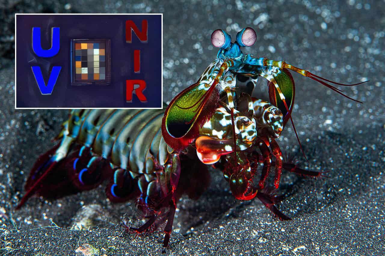
The mantis shrimp prowls the shallow waters of the world’s oceans, armed with some of the most complex eyes in the animal kingdom. Now, that same extraordinary vision is poised to enter the operating theater.
Researchers have engineered a remarkable new camera that mimics the crustacean’s ability to see across multiple wavelengths of light simultaneously. By capturing ultraviolet, visible, and near-infrared light on a single, tiny chip, this bio-inspired device promises to do what current cameras cannot.
The shrimp-inspired vision can show doctors not only where a patient’s lymph nodes are, but whether those nodes harbor hidden cancer cells.
A Surgical Challenge The camera integrates pixel-level filters and stacked light-sensing layers so that ultraviolet, visible and near-infrared signals can be detected separately on one chip. This makes it possible to see both the location and likely cancer status of a lymph node using a compact camera. Credit: Viktor Gruev, University of Illinois at Urbana-Champaign
When a surgeon operates on a patient with breast cancer, the immediate goal is to remove the primary tumor. But the invisible threat lies in the lymphatic system. Lymph nodes act as the body’s defensive filters against cancer cells. They trap rogue cancer cells that travel far away from the main tumor site before those cells can seed themselves elsewhere in the body.
During an operation, surgeons must decide which nodes to biopsy, which to remove entirely, and which to preserve. Remove too few, and the cancer inevitably spreads. Remove too many, and the patient faces a lifetime of painful, debilitating swelling known as lymphedema.
Obviously, high precision in targeting potentially cancerous lymph nodes is preferred. Yet doctors currently lack it. Until now, the standard practice has relied on chemical dyes to simply locate the nodes. Surgeons effectively operate in the dark regarding the actual pathology of the tissue until days later, when a laboratory analyzes the samples.
“Existing tools can show where lymph is draining but can’t reliably indicate whether a particular lymph node is involved with cancer while the operation is underway,” said Viktor Gruev, University of Illinois at Urbana-Champaign.
“This can lead to overtreatment, undertreatment, or the need for a second procedure later.”
×
Thank you! One more thing...Please check your inbox and confirm your subscription.
The Crustacean Eye for CancerHow exactly does a bottom-dwelling crustacean fit into all this? It comes down to photoreceptors.
 Researchers developed a new chip-based camera system to help surgeons identify lymph nodes connected to a tumor while also providing clues as to whether cancer has spread to them. In the image, they are using the system to examine breast cancer tissue. Credit: Viktor Gruev, University of Illinois at Urbana-Champaign
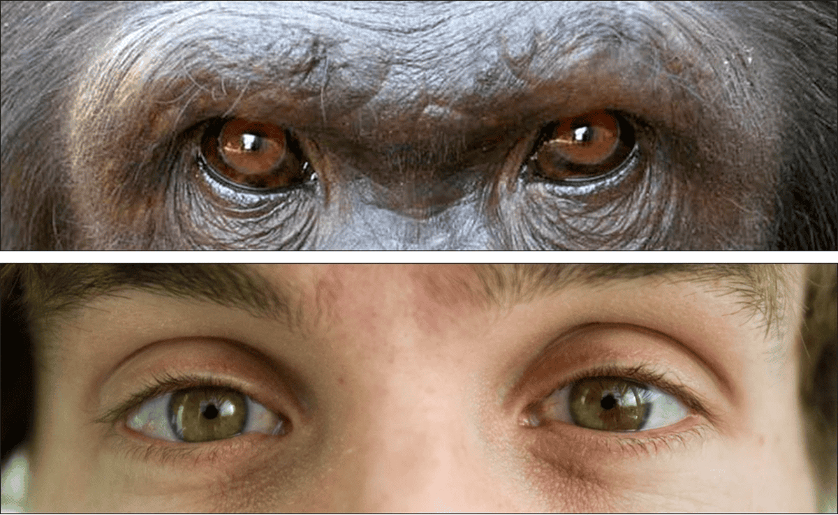
Humans perceive a narrow band of visible light. The mantis shrimp, however, sees much broader. These crustaceans can detect ultraviolet, visible, and near-infrared light because their eyes contain stacked rows of specialized photoreceptors. Each row tunes into a entirely different slice of the electromagnetic spectrum. It’s impossible to truly imagine what a mantis shrimp sees, but sheer number of extra colors must be staggering.
The engineering team looked at this biological marvel and built a digital equivalent. They created a single-chip imager that integrates tiny, pixel-level filters with stacked light-sensing layers.
But if you capture all these different types of light at once, the resulting image could be a blurry mess. To solve this optical traffic jam, the researchers abandoned traditional, bulky glass lenses. Instead, they utilized a specialized mirror-based lens to keep the wide range of wavelengths perfectly in focus together. They then wrote custom image-reconstruction software to weave the disparate signals into a single, razor-sharp picture.
“We borrowed that idea to allow our camera to collect several kinds of optical information from exactly the same place at the same time,” said Viktor Gruev, University of Illinois at Urbana-Champaign.
“This is very helpful when you want images to line up perfectly during surgery.” (Source: Optica press release).
 Researchers have developed a camera inspired by mantis shrimp vision that captures ultraviolet, near-infrared and visible images using a single chip. Shown are single exposures capturing deep UV fluorescence (tryptophan), visible color (Macbeth chart) and NIR fluorescence (ICG). Credit: Viktor Gruev, University of Illinois at Urbana-Champaign
How does seeing different light wavelengths actually spot a tumor? The magic lies in how different biological tissues interact with the spectrum.
First, the surgeon injects a standard fluorescent dye called indocyanine green (ICG). The camera’s near-infrared sensors pick up this dye easily. The dye glows brightly on the surgical monitor, mapping the exact location of the hidden lymph nodes.
Once the surgeon finds the node, the camera deploys its ultraviolet capabilities. When bathed in brief pulses of UV light, cancerous tissue naturally fluoresces differently than healthy tissue. The camera reads this inherent, tell-tale glow. It requires no special cancer-targeting chemicals to do so. Simultaneously, the visible light sensor provides a standard color video feed, giving the surgeon the normal anatomical landmarks they need to navigate the human body safely.
This approach represents a massive leap over previous optical methods. While scientists have known for years that label-free optical techniques can highlight biochemical changes in cancer, previous devices were far too clunky. They required multiple separate cameras and monitors that surgeons could not easily integrate into the fast-paced, high-stakes environment of an operating room. By placing everything on one chip, the researchers have solved the packaging problem.
From the Laboratory to the Operating TheaterTo prove their crustacean-inspired concept, the team put the camera to the ultimate test. They examined freshly removed breast cancer specimens, analyzing 94 distinct lymph nodes from 33 different patients.
Pathologists — the medical detectives who traditionally diagnose cancer under a microscope — reviewed the same tissues without seeing the camera’s readouts. The camera’s ultraviolet sensor identified the cancerous nodes with 97 percent sensitivity and 89 percent specificity. Meanwhile, the near-infrared sensors never failed to map the nodes’ physical locations.
The team now faces the grueling journey from laboratory success to clinical standard. They must test the system on larger, more diverse groups of patients to ensure reliability. They must refine their software to process images even faster in real-time. They also need to ensure the system does not confuse actual cancer with benign conditions, such as harmless tissue inflammation or scarring. Finally, they must package the hardware into a sterile, user-friendly format for daily hospital use.
“If future testing is successful, this system could help patients receive more precise surgery,” said Viktor Gruev, University of Illinois at Urbana-Champaign.
“Beyond breast cancer, the approach could be useful in other cancers where lymph node status matters or anywhere that rapid, label-free tissue assessment could help during surgery or pathology assessment.”
The findings appeared in the journal Optica.


Comments (0)